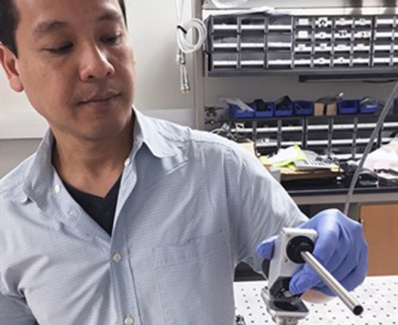
Engineer designs non-invasive device to detect cancerous oral tissue early

Image: Dwight Look College of Engineering
A noninvasive device that enables doctors to quickly and accurately identify cancerous tissue in a person’s mouth could result in more effective diagnosis and treatment of the disease, says a biomedical engineer at Texas A&M University who is developing the instrument.
The potentially life-saving tool makes use of technology known as “fluorescence lifetime imaging (FLIM)” to measure and visualize the biochemical changes that occur in oral epithelial tissue as it turns cancerous, says Javier Jo, associate professor in the university’s Department of Biomedical Engineering. Measuring these specific changes, the technology, Jo says, can assist physicians in differentiating precancerous, cancerous and benign lesions in patient’s mouth.
The research, which is supported by the National Institutes of Health, was presented at this year’s World Molecular Imaging Congress, a venue where scientists and clinicians discuss cutting-edge advances in molecular imaging.
Jo’s device, which is essentially a small, handheld microscope, employs the FLIM technique to noninvasively evaluate tissue for the structural and molecular changes that serve as key indicators in determining if tissue is precancerous or cancerous. With the tool, Jo can observe distinct fluorescence signatures – fingerprints of a sort – that are specific to benign, precancerous and cancerous tissue.
Using FLIM, Jo explains, the fluorescence spectrum (the color content of the fluorescence light) and the fluorescence lifetime (related to the time a molecule emits fluorescence light after being excited) can be detected. Previous implementations of FLIM imaging have been too cumbersome and slow to be used for clinical applications, but Jo’s lab has developed some of the fastest FLIM systems that are being adapted for different clinical diagnosis applications, including oral cancer detection. Another advantage, Jo says, is that there is no need to use any external contrast agent, as only the natural fluorescence of the tissue is being measured.

