

Confocal microscope allows scientists to study near-3D images of tiny, living organisms in real time
Confocal technology is one of the most important advances in optical microscopy, and many disciplines within Texas A&M AgriLife and other parts of The Texas A&M University System are discovering it can also be a game-changer in their research.
Brian Shaw, a professor in the Department of Plant Pathology and Microbiology at the Texas A&M College of Agriculture and Life Sciences, is an expert in confocal microscopy. He not only applies this state-of-the-art technology in his own research, but also encourages undergraduate and graduate students, as well as other researchers, to use it to advance their own scientific efforts.
“Confocal microscopy offers several advantages over conventional microscopy, including the ability to control depth of field and to reduce or eliminate out-of-focus light in image formation on the focal plane,” Shaw explains.
“It also gives you the ability to work with living cells and see the changes they make in real time, as well as to collect serial sections from thick specimens,” he said. “Additionally, it can produce an almost 3-D image of the cell, allowing you to view it from various perspectives.”
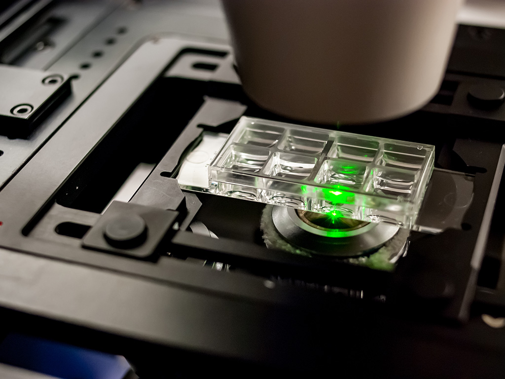
Shaw’s primary confocal microscope, which allows for a wide range of imaging modalities, is in the Plant Pathology and Microbiology Building on the Texas A&M campus.
It is an Olympus FV3000 confocal laser-scanning microscope with four detectors and six laser lines, which allow for simultaneous imaging of four-color channels of almost any fluorophore. It is fully motorized with autofocus, allowing for time-lapsed imaging for hours without losing focus. The microscope’s precision motorized stage makes it possible for multiple-point time-lapse of various specimens simultaneously.
According to research in the Multidisciplinary Journal of Microbial Ecology, more than 80% of crop diseases are caused by fungi or fungus-like pathogens. These diseases lead to billions of dollars in crop losses and threaten food security.
Shaw said much of the research he and his team do relates to determining the cellular machinery involved in the growth and development of fungi and how fungal pathogens work.
“We are investigating surface characteristics of fungal spores and how they influence spore dispersal,” he said. “The fungal spore is the dormant cell that these organisms use to disperse across distance and through time.”
Shaw said fungi require polarized hyphal growth to have an impact on plants and humans, so understanding how hyphae are made is a fundamental concern. A hypha is the basic unit of a filamentous fungus and typically consists of a chain of elongated cells that expand at the apex of the tip cell. The elongated, thread-like cells grow only at their highly polarized apex and their growth is characterized by the initial establishment of one growth site, which is followed by its continuous maintenance.
“Our lab examines the temporal and spatial dynamics of cytoskeletal components during the growth and development of fungal hyphae,” he said. “Using the confocal microscope, we found that the spores of the important corn pathogen, Colletotrichum graminicola, are asymmetric. And these spores can only attach to their new host on one face of the spore.”
Shaw said since attachment to their new host is necessary to begin the disease cycle in corn, this discovery identifies a new and essential target to disrupt fungal disease in corn.