

How does Lyme disease wreak havoc on humans? Interdisciplinary team of Texas A&M scientists looks for answers
Avalon_Studio/istockphoto.com
Researchers at the Texas A&M University’s School of Medicine and its School of Veterinary Medicine and Biomedical Sciences recently published an article in the Journal of Immunology delving into how Lyme disease causes its devastating effects in humans.
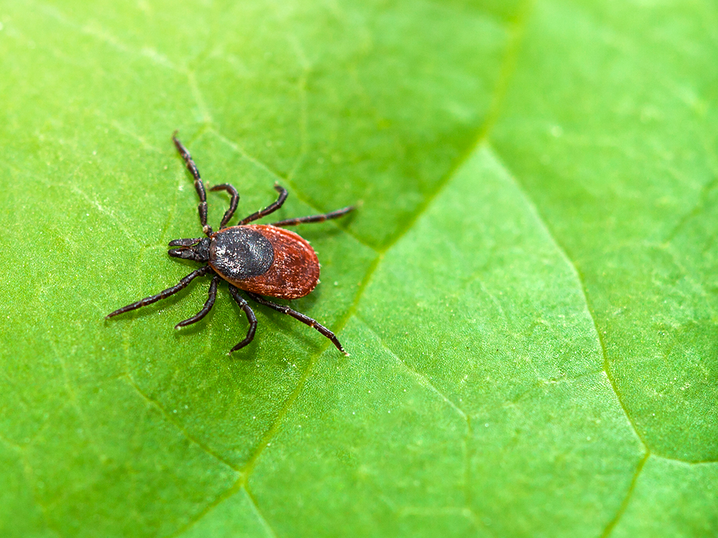
Lyme disease is caused by infection from the bacteria Borrelia burgdorferi (B. burgdorferi), which is carried by ticks. According to recent estimates from the Centers for Disease Control and Prevention, approximately 476,000 patients are treated for Lyme disease each year.
After ticks transmit B. burgdorferi to humans, symptoms can range from fever, headaches, fatigue and a rash to more serious complications occurring in the heart and nervous system. When Lyme disease advances, it can cause chronic fatigue, headaches, muscle pain, irregular heartbeat, neurologic complications and arthritis. These persistent symptoms can arise if the infection was not promptly treated with antibiotics or if the B. burgdorferi bacteria become resistant to treatment. Many chronic symptoms, including joint swelling and arthritis, are directly linked to a strong and lasting immune response resulting from the initial B. burgdorferi infection. Currently, vaccines for prevention or therapeutics to treat chronic Lyme disease symptoms are not available.
B. burgdorferi initiates an inflammatory response in humans by first triggering the innate immune system. The innate immune system responds to infection within a matter of minutes and is built upon the ability to sense molecules shared by many pathogens. In the case of B. burgdorferi, prior research has shown that lipoproteins found in the bacterial cell membrane interact with an innate immune sensor called Toll-like Receptor 2 (TLR2). Although TLR2 is responsible for triggering much of the initial inflammation to infection, other innate immune receptors are likely involved.
Dr. Jenny Hyde and Dr. Phillip West, assistant professors in the Department of Microbial Pathogenesis and Immunology, combined their respective expertise to explore this question in more detail.
B. burgdorferi infection is well-known to engage a family of innate immune proteins called type I interferons (IFN-I). IFN-I is important in the clearance of bacterial and viral pathogens, but also can cause organ injury and chronic disease. Interestingly, IFN-I is heavily linked to Lyme disease-related arthritis and neurologic complications. Given that TLR2 cannot induce IFN-I, Hyde and West postulated that another innate immune sensing pathway must be involved.
Research led by graduate students Lauren Farris and Sylvia Torres-Odio sought to explore the contributions of the cGAS-STING innate immune pathway to IFN-I induction during B. burgdorferi infection. The cyclic GMP-AMP synthase (cGAS)—stimulator of IFN genes (STING), pathway recognizes DNA from viral and bacterial pathogens, but no prior studies had investigated its role in inflammatory and IFN-I responses during B. burgdorferi infection.
As a first step, the research team exposed macrophages and fibroblasts to B. burgdorferi and followed the production of IFN-I and other inflammatory markers. Macrophages engulf bacteria during infection and produce large amounts of inflammatory mediators. Fibroblasts are classically thought of as structural cells; however, the production of IFN-I from fibroblasts has been heavily linked to the development of Lyme-related arthritis. In both cell types, the researchers found that the onset and production of inflammatory compounds began in 24 hours or less.
With these results in hand, the team utilized cells lacking genes that code for the cGAS-STING pathway. When exposed to live B. burgdorferi, macrophages and fibroblasts deficient in either cGAS or STING produced significantly lower amounts of IFN-I. The team also used small molecule inhibitors to block the activity of cGAS and STING and observed reduced IFN-I during infection. Finally, the team utilized microscopy approaches to visualize that the DNA sensor cGAS localizes near internalized B. burgdorferi bacteria, suggesting that cGAS may sense bacterial DNA to engage IFN-I signaling.
The team next examined B. burgdorferi infection in a mouse model using bioluminescent imagining. Here, B. burgdorferi was engineered to express a gene from fireflies that produces light and is detectable with a specialized camera. This technique allowed the researchers to track how much B. burgdorferi was present and where it was located during infection. Interestingly, models lacking cGAS or STING showed no differences in the amount of B. burgdorferi present. However, in collaboration with Dr. L. Garry Adams, senior professor in the Department of Veterinary Pathobiology, the team noted that cGAS deficient models exhibited less joint inflammation and arthritis after B. burgdorferi infection.
Overall, this study reveals that the cGAS-STING innate immune pathway is a critical upstream mediator of IFN-I expression following B. burgdorferi infection.